Sunday 21 March 2021

Myelomeningocele

Myelomeningocele is a condition in which an infant’s spine does not form normally and the baby is born with a fluid filled sac that is visible outside of the back area. This sac containing a part of the spinal cord, its covering called meninges, and spinal fluid, pushes itself through the space between the spine and the skin. Although there is no exact cause for this disease, lack of folic acid, exposure to viruses or radiation, and genetic factors are suspected causal factors. The babies born with this defect have various difficulties like:

  • Weakness and trouble in moving the body parts below the level of the sac
  • Poor bowel and bladder control
  • Excess spinal fluid in the brain (hydrocephalus)
  • Malformed brain
  • Twisted or abnormal lower limbs
  • Impaired cognitive development
  • Seizures

A blood test called the quadruple screen during pregnancy can help detect high levels of a protein called alpha fetoprotein (AFP) in your blood which is suggestive that the baby has a higher risk of developing a myelomeningocele. Other tests like ultrasound  and amniocentesis can help confirm the diagnosis.

While most cases of myelomeningoceles are surgically repaired soon after birth, some cases of myelomeningoceles are treated with surgery while the baby is in the womb prior to delivery. Surgeries are aimed to correct the deformity, limit the risk of infection and prevent more damage to the spinal cord. At Chennai Brain and Spine, our expert neurosurgeons work together involving multiple specialities to cover your baby’s spinal cord safely within 48 hours of birth.


Reviewed By: Dr Shyam Sundar K

Visit Us: chennaibrainandspine.com

Mail Us: shyamsundar_krishnan@yahoo.com

Book Appointment: chennaibrainandspine.com/book-appointment.html

Tuesday 15 December 2020

Spinal Cord Tumors

An abnormal tissue growth within your spinal canal or within the bones of your spine is termed as spinal cord tumor. When the tumor begins from within the spinal cord, it is called intradural tumor and when the tumor affects the bones of the spine, it is called vertebral tumor. Based on the origin of the tumor, it is also classified as:

  • Primary spinal cord tumors:  Tumor that originate in the cells within or next to the spinal cord
  • Secondary spinal cord tumors:  Tumor that originated and spread from another part of the body like the lungs, breasts, thyroid, kidney etc. Secondary tumors are mostly metastatic in nature.

Know the Symptoms

Symptoms are caused due to the pressure on spinal nerve roots. Due to this, the pain may radiate along the course of the nerve, which over a long period of time may cause wasting away of the muscles affected by the nerve. Nerve compression may cause the following symptoms:

  • Severe back pain
  • Decreased sensation, progressive weakness, or paralysis in affected area
  • Loss of bladder and bowel control
  • Erectile dysfunction in men
  • Stiff neck or back
  • Difficulty in walking
  • Decreased sensitivity to pain, heat and cold

Diagnosis

It is very important to diagnose the compression of the spinal cord by a tumor as early as possible for immediate treatment to prevent permanent damage. Diagnosis is done with:

  • Magnetic resonance imaging (MRI)
  • Myelography with computed tomography (CT)
  • Biopsy

Treatment

Spine tumor surgery is vital to restore spinal stability and functioning of the spine. Surgery is also helpful for decompression of neural elements, pain reduction, and preventing the spread of the tumor. Recurrence of the condition can also be reduced to a great extent through surgery. Depending on the condition of each patient the treatment options  like radiation therapy and chemotherapy along with other medications are also combined together for better results. The outcome of the treatment depends on the age, overall health of the patient and on the type of spinal tumor (whether  it is benign or malignant, primary or metastatic). Recovery also depends on how quickly treatment started and how much damage was done to the body. Physical therapy and inpatient rehabilitation will help the patient to restore strength, function and speed up the recovery process.


Reviewed By: Dr Shyam Sundar K

Visit Us: chennaibrainandspine.com

Mail Us: shyamsundar_krishnan@yahoo.com

Book Appointment: chennaibrainandspine.com/book-appointment.html

Tuesday 13 October 2020

Head Injuries - Types, Causes, Treatments

Any injury that causes damage or injury to the scalp, skull or the brain, can be called a head injury. It can be a minor bump or major physical damage. When the brain is affected, it is called Traumatic Brain Injury and can drastically change the physical and mental status of a person. They are also a major cause of death and disability.

Types:

Concussion - where the head is shaken so hard that the brain literally bounces off the side of the skull. This can range from mild to severe. Any sudden deceleration of the body or impact elsewhere can cause the brain to move in the cranium.

Contusion - where a bruise forms on the brain itself, leading to bleeding and swelling.

Intracranial Hematoma - where there is bleeding in the skull that then forms a clot. And, depending on where they form, can range from mild to severe.

Edema - this is swelling to injured tissue. When it happens to the brain tissue, the skull cannot stretch to accommodate the swelling, leading to pressure buildup, pressing the brain against the skull.

Skull Fracture - here the skull bone is fractured and cuts into the brain causing all kinds of complications.

Diffuse Axonal Injury - as there is no visible sign outside, this is one of the most dangerous, often leading to brain damage or death. Here there is damage to the brain cells, but no bleeding. This prevents their function, often leading to more swelling and damage.

Causes:

Vehicular accidents are the most common, followed by accidents at home or workplace and involvement in acts of violence.

Treatments:

This depends on the type and severity of the injury.  Minor injuries may need nothing more than pain medication. But, if there is a cut, it may need to be closed and medicated. Serious injuries may need hospitalisation and the treatment can be:

Medication - as there is a possibility of seizures, anti-seizure medication may be prescribed. Medication to increase the amount of water and salt excretion from the body called Diuretics, may be prescribed. This reduces pressure build up caused by Edema. Sometimes the patient may have to be put into an induced coma, as then the brain needs less oxygen and nutrients.

Surgery - this is an emergency procedure and may have to be carried out to repair physical damage to the skull; release internal buildup of  pressure inside the skull; to remove a hematoma, which is the buildup of blood outside the blood vessels.

Rehabilitation - any serious brain injury could lead to loss of functionality, and regular rehabilitation will be necessary to retrain the brain to normal functioning. What this will be depends on what functionality is lost.

Head injuries are not to be taken lightly and medical help must be sought without any delay.                                    


 Reviewed By: Dr Shyam Sundar K

Visit Us: chennaibrainandspine.com

Mail Us: shyamsundar_krishnan@yahoo.com

Book Appointment: chennaibrainandspine.com/book-appointment.html

Saturday 23 May 2020

Tips to stimulate the brain during a lockdown

The whole world is in the midst of an unprecedented lockdown in response to the Covid-19 pandemic. People everywhere are spending long periods of time indoors, and this is having a detrimental effect on their health, both mental and physical.


Following are some of the steps that we can take to stimulate the brain and hence our mental health.
  1. If you or anyone in your family is on medication, maintain the stock, and make sure it is taken regularly. 
  2. In house exercise routine: a regular exercise routine is not just a way to kill time, but also to stay healthy. A healthy mind in a healthy body. And, if we can make it a family affair, the better it is. The family that exercises together, stays together. The internet is brimming with resources, from old fashioned PT exercises to newer specialised ones. But, do consult with your doctor before you do.
  3. Meditation and/or breathing: meditation works, and has been shown to reduce anxiety, depression and sometimes increase brain volume in specific areas. If that is not your thing, then just breathe. Yes, you already do that but start doing so in a focussed manner. It increases blood oxygen levels and reduces stress levels.
  4. Maintain or renew social or community connections: take that phone, call your friends, family, neighbours. Check up on their affairs, connect with them. We are social animals, we need this connection. Make it; keep it.
  5. Brain exercise: Anything that gives your brain a workout is acceptable. Puzzles of all kinds; chess, Go, knowledge quizzes. It is not the winning, but the playing, the together time, that matters.

Remember, the pandemic is not going anywhere soon. But, that does not mean we should give up.  Stay healthy, stay safe.


Reviewed By: Dr Shyam Sundar K
Visit Us: chennaibrainandspine.com
Mail Us: shyamsundar_krishnan@yahoo.com
Book Appointment: chennaibrainandspine.com/book-appointment.html

Tuesday 25 February 2020

Awake craniotomy: What you need to know?

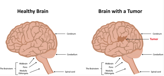
Awake craniotomy is a type of brain surgery, during which the patient remains awake. This procedure is carried out to manage cancer of the brain and skull base. A general anesthetic is used to minimize discomfort during the procedure.

 Brain tumor

Why is it done?

Awake craniotomy is usually recommended when a brain tumor involves or is close to parts of the brain that control vision, movement or speech and that must be preserved. This can avoid any potentially life-changing damage during the surgery. 

Why is the patient kept awake?

Awake craniotomy is often opted, when the Functional MRI can not pinpoint the most critical areas of the brain which are to be preserved. These vital areas are located using special speech mapping techniques while the patient is awake in the operating room. Awake speech mapping is carried out by applying a mild electrical current to the surface of the exposed brain. 

The patient will be asked to perform various tasks, such as reading or talking or blinking or moving fingers. Surgeons will monitor reflexes and reactions and when stimulation hinders the task, then that area of the brain is marked and preserved. When the stimulation blocks the task, it is considered as the area of the brain that is marked and preserved. This can minimize the complications impacting the patient’s sight, speech or movement. 

How is the surgery performed?

Patients will receive general anesthesia before surgery. This will keep them relaxed and calm throughout the procedure. The surgeons will make an incision and remove a small portion of the skull and start awake speech mapping. According to the stimulation, the surgeons will mark and protect critical areas of the brain that control important functions. Awake craniotomy usually lasts four to six hours. After which the patient will spend one to three days in observation, depending on the condition return to normal activity within one to two weeks. 

Is it painful?

Awake craniotomy is not painful. As mentioned earlier, the patient will receive general anesthesia and they don’t feel any discomfort. One interesting fact is that the brain doesn’t have any pain receptors so the surgeon can operate without risk of causing pain.  

Just like every other brain surgery an awake craniotomy is a highly complex procedure. However, compared to other brain surgeries awake craniotomy facilitates safe and faster recovery. Most often, 75 percent of patients can go home 24 hours or less after surgery. 

 Brain surgery

Book an appointment at Chennai brain and spine for expert guidance and discuss with expert surgical oncologists and neurosurgeons who have specific experience in removing cancers from high-risk areas of the brain.

Reviewed By: Dr Shyam Sundar K
Visit Us: chennaibrainandspine.com
Mail Us: shyamsundar_krishnan@yahoo.com
Book Appointment: chennaibrainandspine.com/book-appointment.html

Tuesday 17 December 2019

Recovery after endoscopic skull base surgery

Endoscopic skull base surgery is the surgery to treat the disease like the tumor or an infection in your brain. This surgery involves open brain surgery to treat.


Complications you may experience after endoscopic skull base surgery:
  • You may also have headaches or problems concentrating. It can take up to 4 to 8 weeks to recover from surgery.
  • Your wound may be painful after surgery for a minimum of 5 days. 
  • You might experience shooting pains and numbness near your incisions, or bruising and swelling may appear around your eyes. 
  • The stitches of your wounds go away on their own or need to be removed by doctors within 7 to 10 days, depending on the stitches type.
Here are the following steps to recovery after endoscopic skull base surgery:
  •  Getting enough sleep will help you recover.
  •  Avoid resting or sleeping in a flat position. You can also raise the headboard of your bed or use a wedge pillow. It prevents headaches or dizziness.
  •  Start walking every day because it boosts the flow of blood and prevents constipation and pneumonia.
  •  Avoid heavy lifting or exercising until your doctors give you heads up.


Diet
  •  There is no particular diet; you can maintain a regular diet. However, when you get bad feelings in the stomach, try low-fat foods or bland, like boiled chicken, toast, plain rice, and yogurt.
  •  Fluid intake should be under a doctor’s recommendation.
  •  Avoid drinking alcohol during the recovery procedure.
Medicines
  • Take medicines only prescribed by doctors.  Also, you need to complete the full   course of medicines as prescribed by your surgeon.
  •  Keep the wound clean and dry. Change the bandage as directed by your doctor.
  •  Follow-up with your doctor regularly and take only prescribed medicines. Also, do not exceed or suppress the dosage of your medicine.
  •  Call your doctor if you are having any problems even unrelated to your surgery. 
This is a general idea about recovery. However, each person recovers at their pace.


Reviewed By: Dr Shyam Sundar K
Visit Us: chennaibrainandspine.com
Mail Us: shyamsundar_krishnan@yahoo.com
Book Appointment: chennaibrainandspine.com/book-appointment.html

Monday 21 October 2019

What Causes Craniocervical Instability

The craniocervical junction is the place where the skull and the upper spinal cord meet. Craniocervical instability, also known as the syndrome of Occipitoatlantialaxial Hypermobility, is a pathological condition that causes structural weakness in the craniocervical junction. This instability might lead to the deformation of the upper spinal cord, brain stem, and cerebellum.
https://www.chennaibrainandspine.com/craniocervical-junction-abnormalities.html

CAUSES

The different reasons for craniocervical instability to occur might include:

1.Ehlers-danlos Syndrome – This is the syndrome that affects the connective tissues in the body. This condition loosens the ligaments and muscles that hold the parts of the body. It is one of the primary reasons that might lead to craniocervical instability. It is estimated that 1 in 15 people suffering from Ehlers-Danlons syndrome will develop craniocervical instability.

2.Physical Trauma – Any form of physical injuries like accidents, neck injury, or blow to the head that affects the connecting nerves or ligaments can lead to the development of craniocervical instability.

3.Neoplasms – Abnormal growth of cells(tumors) that do not promote the growth of other cells are called neoplasms. Conditions like a bone cyst or blood vessels clot near the cranial nerves might lead to cervical instability.

4.Inflammatory Diseases – Conditions like arthritis that cause swelling or inflammation of the joints might also be one of the reasons for the development of the condition.

5. Congenital– the diseases like downs syndrome or dwarfism that are present from the birth might also cause instability in the craniocervical junction.

6.Nerve Dysfunction – repetitive stress or stretching that occurs on the craniocervical nerves might lead to instability.


7. Cranial Settling – It is the condition when the skull sinks downward on the spine, causing pressure on the surrounding nerves and muscles. The pressure exerted might lead to instability in the cervical region.

8.Retro Flexed Odontoid – Misaligned odontoid bone can be caused due to loose ligaments that push backward, which in turn compresses the brain stem causing instability.

Craniocervical instability can be identified with signs such as severe headaches, pressure headaches, neck pain, facial pain, muscle weakness, sleep apnea, and many more.

Usually, the condition is diagnosed using MRI and CT scans or by Invasive Cervical Traction. The most common treatment method is to perform a craniocervical fusion.

For any neurological related problems, contact Dr.Shyam Sundar K, One of the leading neurologists based in Chennai.
https://www.chennaibrainandspine.com/contact-neurology-doctor.html

Reviewed By: Dr Shyam Sundar K
Visit Us: chennaibrainandspine.com
Mail Us: shyamsundar_krishnan@yahoo.com
Book Appointment: chennaibrainandspine.com/book-appointment